|
Acarine
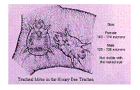
Cause Acarine, or more properly acariosis, disease is caused by the Acarapis woodi mite. This is a small mite about 150 X 65 μ which lives and breeds in the tracheal tubes of the adult bee. Disease The entire life cycle of this mite is spent within the respiratory (tracheal) system of the honey bee, except for brief migratory periods. Within 24 hours after worker bees emerge from their cells as new adults, female mites collect within their tracheae, where the mite feeds and reproduces. Mites also may be found in air sacs in the thorax, abdomen, and head. The mites pierce the breathing tube walls with their mouth parts and feed on the haemolymph, or blood, of the bees. As a result of mite feeding, the haemolymph of infested bees has a higher than normal bacterial count. Each female mite lays five to seven eggs, which require 3 to 4 days to hatch. Male and female mites develop from egg to adult in approximately 11 to 15 days. Eggs hatch into six-legged larvae, then moult to a non-feeding nymph stage, and then finally moult to the adult stage. All stages of the mite - eggs, larvae, and adults - may be found in the tracheae of older bees. Signs in the colony This disease was once called Most sources now do not associate any specific signs with acariosis. There is no known link between acariosis and CBPV. It was more conviction by association. It is possible however that the mite may stress the bees sufficiently to let the viruses cause disease. As mite populations increase, colony populations dwindle and this can ultimately result in the death of the colony. Colonies are most affected during winter confinement and early spring like a stress disease. Mite infestations are at a maximum in the late winter and early spring when the population is composed of primarily older bees.
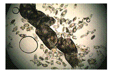
Positive identification of tracheal mites can be done only by dissection and microscopic examination of honey bee thoracic tracheae. The tracheae of uninfested bees are clear and colourless or pale amber in colour (healthy). In a slight infestation, one or both tracheal tubes contain a few adult mites and eggs, which may be detected near the spiracle openings. At this stage, the infested tracheae may appear clear, cloudy, or slightly discoloured. Infested tracheae undergo progressive deterioration and show patchy discoloration. The tracheae of severely-infested bees have brown blotches with brown scabs or crust-like lesions, or may appear completely black, and are obstructed by numerous mites in different stages of development. Feeding by the mites damages the walls of the tracheae. The bee's flight muscles (in the thorax) may also become atrophied in severe infestations Treatment There is no approved treatment for the mite in the It is likely that treatment for Varroa with pyrethroids will reduce the numbers of acarine mites at the same time. Control Spread of the mite is by direct bee contact. Taking steps to prevent robbing and drifting will reduce the spread between colonies. As there is no approved treatment maintaining the colony in a stress free environment will reduce the risk of secondary disease. If the colony becomes severely infested it should be
destroyed. |
 Diagnosis