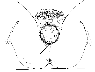
This begins at the mid-point of the fourchette and continues at a 45 degree angle to the midline (towards 7 o' clock). This incision reduces danger of damage to the anal sphincter and Bartholin's gland. It is the most common incision used in the UK.(ref 4)
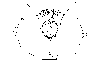
This is a mid-line incision. It is associated with reduced blood loss, less pain and improved cosmetic appearance, but there is a high risk of damage to the anal sphincter if the incision tears further.
Spontaneous trauma is classified according to the structures involved. This ranges from first degree tears, which involve the posterior wall and perineal tissue only to third degree tears which include the anal sphincter and longitudinal muscle.
The risk with the lateral incision is that the laevator anai muscle might be weakened and that the Batholin's Gland may be severed
Suturing a J-shaped incision is very difficult and the repair tends to be puckered

The instrument trolley should be set out with
- A needle holder
- Scissors
- Small artery forceps
- T forceps
- Cotton wall balls soaked in antisceptic solution
- Swabs
- Needle disposal pad
- Tampon
- Sutures
All swabs and instruments should be counted before and after the procedure.
Ensure that the local anaesthetic is well inside its expiry date, explain the procedure to the woman before placing her in the lathotomy position with her legs in stirrups
The vulva is swabbed with an antiseptic solution from anterior to posterior direction. A tampon may be inserted to stem the uterine blood flow. A small artery forcep should be attached to the tail. A thorough examination of the vagina and labia should be carried out to identify the extent of the trauma. If you are at all concerned, then a doctor should be informed, particularly if the anal sphincter or muscles are involved in the tear. Local anaesthetic (amount and strength depending upon unit policy) is infiltrated into vagina, muscle and skin edge. The full length of the needle is inserted into the tissue, ensuring that it has not entered a blood vessel and then the local anaesthetic is inserted as the needle is withdrawn. This is infiltrated into skin edges in a kite shape to ensure even distribution. Check that area is adequately anaesthetised before suturing commences.