|
|
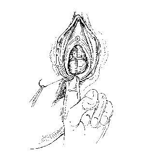
The apex of the incision is identified
at the uppermost limit of the wound in the posterior wall
of the vagina. The first stitch must be inserted above
this point in order to secure any bleeding points which
may have retracted beyond the skin edges. The first
stitch is secured, taking care not to tie the knot too
tightly. Oedema will develop during the first 24-48 hours
and sutures which have been tied too tightly will
constrict the tissue, causing pain and compromising the
repair. The posterior vaginal wall is now closed using a
continuous locking stitch, which ensures good
haemostasis. Take bites of vaginal skin of approximately
on either side. The hymnal remnants are useful landmarks
here and these should be brought together carefully. It
is important to identify the fourchette and to ensure
correct apposition at this point. When the fourchette is
reached insert the needle through the vaginal wall and
bring it out into the muscle layer. This first muscle
stitch finishes off the vaginal repair. Now check that
haemostasis has been achieved. |
|
|
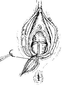
The next step is the repair of the
perineal muscles. Before commencing the muscle layer,
check the depth. It may be necessary to insert two
layers, but normally one layer of interrupted sutures is
sufficient. At the apex of the tunnel created by the
vaginal skin closure a bite of muscle is taken on either
side using the same needle as was used for the vaginal
repair. This first muscle stitch finishes off the vaginal
repair. Knots can either be tied superficially or buried.
Either way, the sutures are brought out close to the skin
surface so that the skin edges can be brought together
without any tension. When closing the muscle layer it is
important to close off all the dead space which could
otherwise lead to a haematoma forming. Care should also
be taken not to include the rectum or anal canal as this
could result in a fistula forming. |
|
|
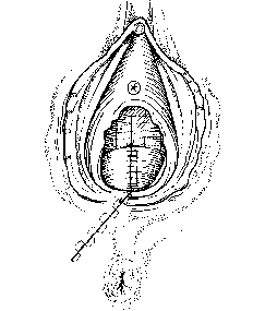
The final step is to close the skin
incision. Studies (Ref 5) have shown
that this method is associated with fewer short-term
problems such as pain on sitting and walking and
dyspareunia. Start at the inferior end of the wound
and take a small bite of subcuticular tissue or a small
bite on either side before tying the first knot. The
short end can then be cut very close to the knot. Reverse
the needle and continue taking small bites on alternate
sides. bringing the edges of the skin together without
any tension. The sutures should be placed deeply in the
subcutaneous layer with a resulting tissue separation of
2-3mm between the skin edges on completion. When the
fourchette is reached reverse the needle so that the knot
will come to lie within the suture line. Keep one end of
the suture as a loop in order to tie off the knot. Tie
the first throw, then reverse the second throw. Add a
third throw for security and cut off the loop only. The
needle is then taken back into the vagina to invert the
knot and bury it deep within the suture line.
|
| |
|
| |
|
Alternatively the skin layer can be closed using interrupted
stitches. There are several ways of tying the knots - they can be
either buried or left on the surface. The mattress stitch has the
advantage that you can control the tightness of each stitch and
they are easy to remove if necessary. Take a large bite of about
5mm on either side of the wound, then reverse the needle and take
a smaller bite of about 2mm on either side. The suture is then
tied as before and the ends are cut leaving long tails to
facilitate removal. Continue until the wound edges have been
brought together. Once the wound has healed the knots can be cut
if required. If absorbable sutures -such as VICRYL or Coated
VICRYL - have been used there is no need to remove the buried
material, as this will gradually absorb.
When the suturing has been completed, the tampon is removed.
Check that haemostasis has been achieved throughout. The vulva is
swabbed and the rectum is checked digitally to make sure that no
sutures have penetrated. Should this be the case, a doctor must
be informed immediately. Swabs, needles and instruments must be
rechecked immediately after the procedure. The area is cleaned
and a sanitary pad placed over the vulva and perineum. Remove the
mother's legs gently and simultaneously from the lithotomy
support and make sure she is comfortable. A record should be kept
of details of the repair, including information about the
perineal infiltration, the indication for performing the
episiotomy and any details of the repair, including the type of
sutures used and the number to be removed (if any).
References
- Grant A. Repair of perineal trauma
after childbirth. In Effective Care in Pregnancy and
Childbirth, Editors: Chaliners I, F.nkin M. Keirse
M. Published by University Oxford Press. 1989 printed by
permission of Oxford University Press.
- Gemynthe et al New VICRLY*
formulation: an improved method of perineal repair. British
Journal of Midwifery, May 1996, Vol 4 No 5.
- Grant A. Repair of perineal trauma
after childbirth. In Effective Care in Pregnancy and
Childbirth, Editors: Chaliners I, F.nkin M. Keirse
M. Published by University Oxford Press. 1989 printed by
permission of Oxford University Press.
- Myles. Textbook for Midwives
Churchill Livingstone 9th Edition p.612
- Grant, A. The choice of suture
materials for repair of perineal traums: an overview of
the evidence from controlled trials. British Journal
of Obstetrics and gynaecology 1986. vol 93, pp 417 -
419