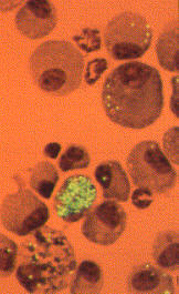
Above: Inert green fluorescent microspheres engulfed by motile lung macrophages.
Lung macrophage dynamics can be quantified by flow cytometry or confocal microscopy. |
Fluorescence microscopy |
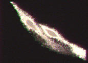
The Magiscan TARDIS image analysis system for the in vivo dynamic video imaging of fluorescent chemical probes within living cells |
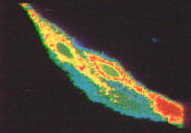
Above: Calcium transients within a bone osteoblast cell using the chemical probe
FURA-2. Where blue = low Ca levels, and red = high Ca levels. Similar techniques
are used to measure intra-cellular pH in lung macrophages using BCECF chemical
probes. |
Return to Home Page |
Return to Index Page |