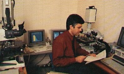
Magiscan image analyser in 1992 Dr. Keith Morris's system installed at UKAEA Harwell. |
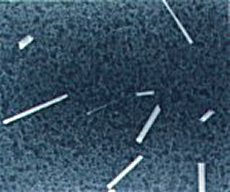
Asbestos bodies These asbestos fibres were recovered from human lung at post-mortem and are evidence of exposure to asbestos during life. |


Above: Scanning electron micrograph of glass fibres recovered from the lung. These fibres were manufactured by Owens Corning Fiberglas with a uniform diameter of 2 um. The fibres were used to test the in vivo lung durability of glass fibres. Durabilty in the lung is an important aspect of the potential hazard from accidental exposure, the others being dose and fibre dimensions. Left: Histogram of the fibres length and diameter distributions These were obtained by phase contrast optical microscopy using the Magiscan image analyser. |
This work investigates the potential hazard from the inhalation of man made vitreous
fibres (MMVF), such as glassfibre, Rockwool and ceramic fibres. Given the proven
hazard of asbestos fibre exposure there is concern over the safety of these
materials. |
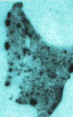
Right: An autoradiograph of an entire lung lobe The lung contained inhaled radioactive asbestos fibres. Before inhalation, constituent elements within the asbestos had been neutron activated inside a nuclear reactor. This radioactivity could then be detected by photographic autoradiograph techniques to show the location of the asbestos fibre within the lung section (dark areas indicate high asbestos burdens). This is a specialised biological radiotracer technique that can measure asbestos and MMVF fibre burdens in vivo. |
Once inside the body radioactive particles can irradiate cells within body tissues.
Such tissue irradiation can cause cancer by damaging stem cells in the somatic
tissues or can cause inheritable genetic damage to offspring by damaging the
immortal germ cell line within the testes and ovaries. After some time post-exposure
the radioactive material may be expelled from the body via physical
lung clearance mechanisms or the urine/faeces. However it can also
relocate to other locations within the body by physical transport (e.g. in motile
macrohage cells) or by particle dissolution. It is therefore necessary to
undertake long term studies to get a clear picture of total lifetime dose resulting
from exposure. |
CR-39 plastic autoradiograph |
H&E stained lung serial tissue section |
This type of work investigates the dosimetric implications of the inhalation or ingestion
of actinide materials such as plutonium and uranium dioxide. Owing to
their largescale use in the nuclear and defence industries these materials are
now a serious hazard to workers and the general public. Plutonium-238 is one of
the most toxic substances known to man, and only certain nerve agents are more
toxic per unit mass.These radioactive substances emit alpha particles, which,
although having a short range, are extremely hazardous if the material is taken
into the body. |
Left: K5 liquid emulsion autoradiograph of plutonium dioxide particles in lung tissue This section contains inhaled insoluble alpha-emitting plutonium-239 dioxide particles. The alpha particles leave black 'tracks' forming 'stars' within K5 photographic emulsion. Again the number of 'tracks' relates to the mass of plutonium in the lung tissue. |
The number of 'tracks' counted in different tissue regions is directly related the
mass, and hence radioactivety, of the actinide particles located there. The volume
of tissue can be calculated from tissue section thickness and 2D surface
area. This information can be used to estimate the dose to different tissue regions
at any given time post exposure, i.e. from the radioactivity per unit mass
of tissue. |
Some Research Interests Dr. Keith J. Morris |
Fibre Toxicology |
Lung Micro-Dosimetry |
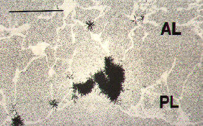
Above: High magnification of CR-39 plastic autoradiograph used to locate uranium
dioxide particles in the lung The black 'stars' are made up of neutron-induced uranium-235 fission fragment 'tracks' from inhaled enriched uranium particles which are etched into the CR-39 plastic along with a shadow image of the lung tissue. The highly insoluble uranium dioxide particles are mainly within motile alveolar macrophage cells that scavange within the alveolar (AL) lung tissue, in this case congregating to form a large cluster at the lung periphery (pleura: PL). |
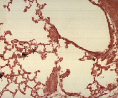
Below: CR-39 plastic autoradiograph used to locate uranium dioxide particles deposited
within lung tissue The uranium dioxide particles are denoted as a series of uranium-235 neutron-induced fission fragment 'tracks' (black stars) eminating from the particle. The number of 'tracks' is proportional to the mass of the particle. Below the CR-39 autoradiograph is a standard serial H&E stained section of the same location showing the detailed tissue structure. |
Return to Home Page |
Plus brief details of my work with fluorescence microscopy, the in-vivo dynamic video imaging of cells, bone morphometry and osteoporosis. |
Secondary ion mass spectrometry |
Further information on biological elemental microanalysis using Secondary Ion Mass
Spectrometry (SIMS) - data from 1993 |
Air pollution research |
A few details of my PhD research in atmospheric aerosol science at the University
of Leeds -1981 |
Short preview of chapter download (Introduction only) - 1995 |
Bioaerosol full chapter download File: bio.doc (as bio.zip) Download of full chapter and figures in a Word 2000 format compressed Zip file. Use an UnZip utility such as TurboZIP Express Uncompressed = 5 Mb, Compressed zip = 1.2 Mb |
The detection and identification of airborne particles of biological origin
that have implications for health and air pollution. Chapter title: Modern microscopic methods of bioaerosol analysis |
Air pollution research |
Image analysers |
Image analysis Details of the modern image analyser |
Index Page |
Dept of Cell Biology |
Cell Biology Divisions imaging suite at the Institute of Ophthalmology, UCL - 2002 |