![Go to Carl Zeiss Microscience [Bio-Rad confocal]](biorad_icu_259x382.jpg)
![Go to Carl Zeiss Microscience [Bio-Rad confocal]](biorad_microscope_289x399.jpg)

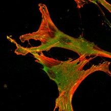
Above: actin cytoskeleton (red) and vinculin (green) at substrate attachment points
in primary human corneal fibroblasts |
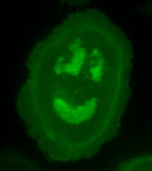
Above: cell expressing GFP tagged EGF receptor |
Above: Double immunoflourescence of subconfluent MDCK cells stained with mAb TJ3/36
and anti-vimentin antibodies or TRITC-phalloidin |
Above: Time lapse images showing annexin2 comet trails |
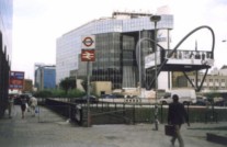
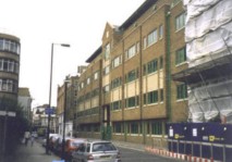
Division of Cell Biology Institute of Ophthalmology University College London 11-43 Bath Street London EC1V 9EL |
Left: The institute buildings Below: Old Street station |
Dr Keith J. Morris in the Cell Biology imaging suite, using the Improvision OpenLabs
imaging workstation |
Above: The evanescent wave microscope under construction with its 35kW SP Beamlok
laser |
Above: the Bio-Rad Radiance 2000 confocal microscope with Zeiss Axiovert 100 microscope |
Left:: Improvision OpenLabs imaging time-lapse system |
Return to Home Page |
Visit Cell Biology's Imaging Facilities Webpage |