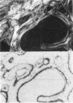
Bone density & histomorphometry |
Above: Cancellous bone section viewed under polarised light (top) and Pu alpha-particle
tracks in a CR-39 autoradiograph of the same bone region (below) |
Quantitative image analysis for measuring bone density and morphometry is performed
using the Magiscan image analyser |
Return to Index Page |
Return to Home Page |
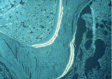
Below: Tetracycline labeling (bright area) for determining bone turnover |
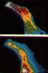
Above: Two SIMS maps showing calcium (top) and phosphorus concentrations (below) in a single
trabecula of cancellous bone |