A brief history of some other image analysers managed by Dr Keith Morris from
1975 to 1990 |
Return to Index Page |
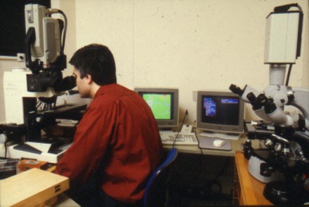
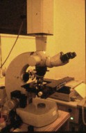
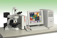
The Joyce Loebl Magiscan With four full RGB colour inputs and 40Mb of dedicated fast video RAM and three large boxes of dedicated hardware, the Magiscan was one of the top machines of its day. It cost over Ł100,000 with a software driven Ł18,000 eight slide motorised stage attached to a Ł40,000 Nikon Microphot mono objective microscope with autofocus and plan optics (1990 prices). The Sony 3 CCD RGB camera shown left cost nearly Ł10,000. On the right is a Bosch Newvicon B&W tube camera attached to a Zeiss Universal photomicroscope with stand alone motorised stage and controller (shown below right). |
Despite its age, the Magiscan will still undertake image analysis operations faster
than many modern cheaper machines owing to its high poportion of dedicated hardware.
The GENIAS software ran on a 286 PC linked to the image processing,
video capture and stage controller modules. The PC was later upgraded to a
486 to help the RESULTS data handling and associated graphics software run faster,
although this had no effect on image processing speed. This Magiscan Colour
was the last model built by Joyce Loebl (Applied Imaging USA purchased the company
in the 1990's and discontinued production and development of general purpose
image analysers, concentrating on cytogenetic applications instead). The Magiscan Colour also ran dedicated software such as FIBRE for measuring 'curly' fibre size distributions, GEMINI for 2D gels, TARDIS for dynamic video imaging of intracellular probes, BONE for bone tissue morphometric and densimetric analysis, plus cytogenetic metaphase finder and chromosome karytyping software. For a clear description of the operation and use of image analysers with various microscopes (optical, confocal and electron) you are referred to chapter 10 in the Bioaerosols Handbook. |
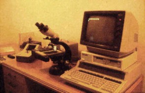
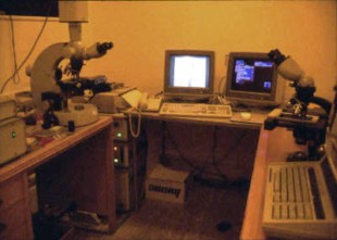
The Magiscan image analyser prior to the installation of the Nikon Microphot with
motorised 8 slide stage. The Magiscan control modules can be seen stacked on the
floor to the left of the grey desk. The Magiscan has a resolution of 512 x 512
pixels, ideal for interactive use as you can actually see the pixels on the
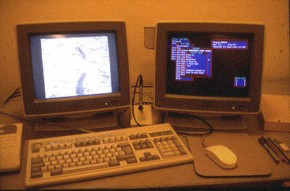
screen for editing. Below left: One VDU is dedicated to displaying the captured or real time image for thresholding and editing via a dedicated light pen. The right VDU is attached to the IBM PC and runs the image analysis controlling software, such as GENIAS, RESULTS or FIBRE. |
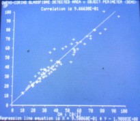
Left: Graphical output from the Magiscan RESULTS package. This program accessed the
binary DAT files created by the image analyser. Being DOS based this is more
complicated at transfering data to other software, compared to a Windows XP based
image analysis system such as the Leica Q550 shown below right. However being DOS based it processed raw data quite fast. |
Above: Fortran 77 data handling and control programs, adapted from those developed for the Seescan system |
Above: Leica Quantimet Q550 |
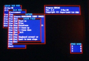
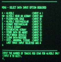
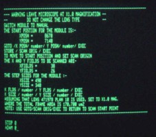
Above: The GENIAS menu running in interactive mode. All the Magiscan image analysis
or results software could be run from batch files or Pascal programmes for fully
automatic or semi-automatic operation. |
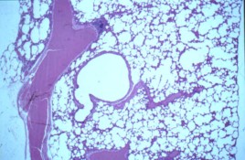
Left: An H&E stained lung section for lung morphometry and particle distribution
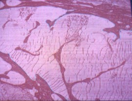
studies Below: A CR-39 autoradiograph of a serial lung section |
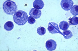
Above: A Shandon Scientific Cytospin of macrophages recovered from a lung after exposure
to glass fibres |
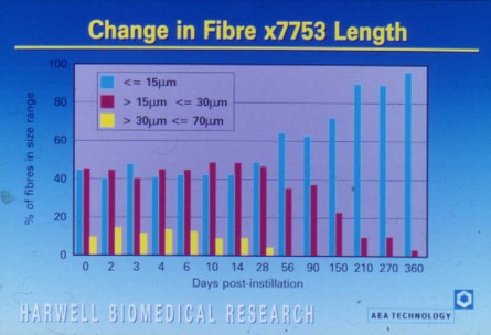
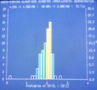
Below: Results from an Owens Corning Fiberglas study of the dissolution of insulation
glassfibres in the lung. Note that all the long fibres dissolve after 28 days
in the lung while the shorter ones (<15 µm long) are protected from dissolution
by being held within macrophages at a different intracellular pH |
A gallery of related topics and pictures including SEM and Flow Cytometry at Biomedical
Research |