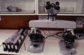
Zeiss bridge micoscope |
H&E |
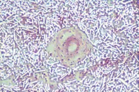
CR-39 alpha-image |
H&E |
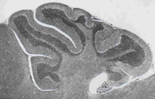
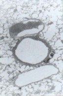
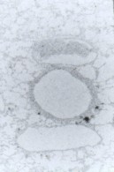
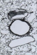
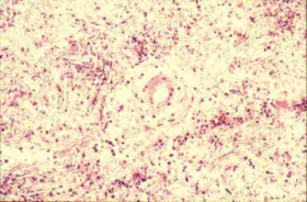
Brain |
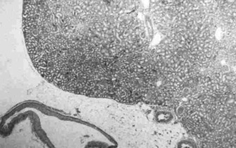
Kidney |
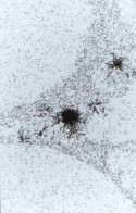
Boronated tissue n-alpha images |
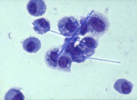
Cytospin |
Phase contrast |
H&E |
SEM electron probe elemental analysis |
SEM |
Above: Lavaged alveolar macrophages recovered from the lung after the inhalation
of fibres |
H&E |
Return to Home Page |
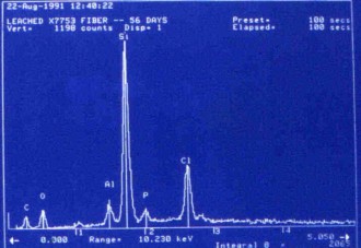
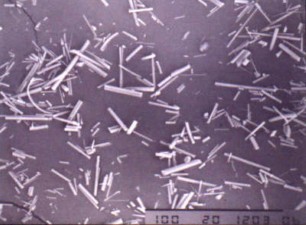
An SEM photomicrograph of glass fibres prior to inhalation |
Elemental analysis of these fibres after 56 days in the lung |
Images of brain and kidney tissue made in CR-39 plastic slides using the boron-10
n,alpha reaction |
Particles of uranium dioxide in the process of being cleared from the lungs by alveolar
macrophages |
The visualisation of glass fibres on intra-peritoneal tissue using phase contrast
optical microscopy |
The use of Boron -10 and various atomic reactor neutron fluences to produce images
of lung tissue and inhaled uranium-235 fission fragment tracks without destroying
the original tissue section (seen right in b&w) |
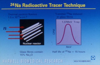
Above: A radioactive in vivo tracer technique for measuring the dose and initial clearance of inhaled glass fibres
using the isotope sodium-24 |
Gallery |
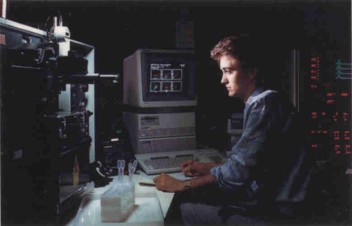
The FACStar Flow Cytometer |
The Biomedical Research high resolution gamma scintillation counter |
Another manual gamma scintillation counter with heavy lead shielding |
Fused aluminosilicate clay particle (FAP) production for lung distribution studies |
Biomedical's histology laboratory |
Return to the Magiscan Page |
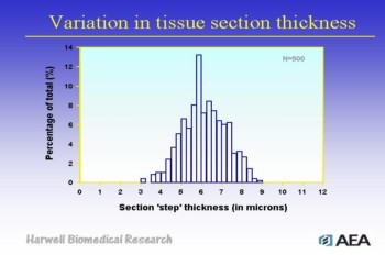
Above: Investigating the variation in the thickness of resin embedded histological
sections cut to a nominal thickness of 5µm (n=500) |
High mag |